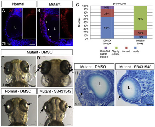
TGFβ signaling drives formation of cellular masses in plod3-mutant lenses. (A,B) Single confocal planes of eyes from 75 hpf normal (A) and plod3vu222/vu222(B) embryos labeled for pSmad3 (red) and nuclei (DAPI, blue). Numbers of eyes analyzed: A = 8, B = 10. Arrowheads in B point at the abnormal ALE that shows expression of pSmad3, and insets in A and B show pSmad3 channel alone in the ALE region. (C–F) Live 4 dpf plod3vu222/vu222 embryos (C,D,F) or normal sibling (E), dorsal views, anterior to the top. Embryos in C,D,E were treated from 30 hpf with vehicle (DMSO) and in F with SB-431542. Arrows point at lenses. (G) Quantification and statistical analysis of treatment of plod3vu222/vu222 with SB-431542 (χ2 test). Location of lenses relative to retina is represented by different colors. (H,I) Transverse histological sections from eyes of plod3vu222/vu222 embryos at 4 dpf. Embryo in H was treated with vehicle and in I with SB-431542. Ten eyes were analyzed in two separate experiments. Arrows in H point at cellular mass and in I at rescued monolayer of ALE with abnormal cells. Scale bars are 20 μm (A,B,H,I) or 100 μm (C–F).
|
