FIGURE
            Fig. 1
                
                    
                        Fig. 1


| 


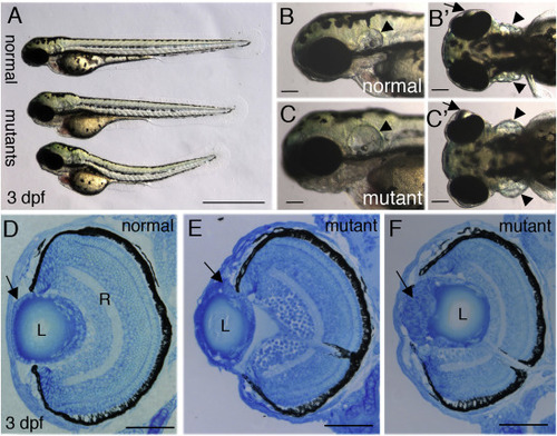
 pninavu222 mutant embryos. (A-C’) Live 3 dpf normal and mutant embryos. (A) Lateral view, top embryo is normal, bottom two embryos are mutants (n > 100 embryos). (B,B’,C,C’) Higher magnifications of head region, lateral views (B,C) and dorsal views (B’,C’) of normal (B,B’) and mutant embryos (C,C’). Arrows point at lenses, arrowheads at ears. (D–F) Transverse sections of eyes from normal (D) and mutant (E,F) embryos at 3 dpf (n > 10 for each genotype). Arrow in D points at the ALE and in E,F at abnormal masses of cells. L, lens; R, retina. Scale bars are 1 mm (A) and 50 μm in all other panels.  | 


                        Expression Data


                        Expression Detail


                        Antibody Labeling


                        Phenotype Data


    | Fish: | |
|---|---|
| Observed In: | |
| Stage: | Protruding-mouth | 
                
                    
                        Phenotype Detail


                        Acknowledgments


This image is the copyrighted work of the attributed author or publisher, and
ZFIN has permission only to display this image to its users.
Additional permissions should be obtained from the applicable author or publisher of the image.
    Reprinted from Developmental Biology, 458(2), Taler, K., Weiss, O., Rotem, S., Rubinstein, A.M., Seritrakul, P., Gross, J.M., Inbal, A., Lysyl hydroxylase 3 is required for normal lens capsule formation and maintenance of lens epithelium integrity and fate, 177-188, Copyright (2019) with permission from Elsevier. Full text @ Dev. Biol.