
Fig. 7
- ID
- ZDB-IMAGE-200413-38
- Antibodies
- Publication
- Taler et al., 2019 - Lysyl hydroxylase 3 is required for normal lens capsule formation and maintenance of lens epithelium integrity and fate
- All Figures
- Figures for Taler et al., 2019
|
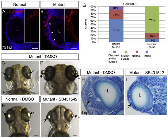
Fig. 7 TGFβ signaling drives formation of cellular masses in plod3-mutant lenses. (A,B) Single confocal planes of eyes from 75 hpf normal (A) and plod3vu222/vu222(B) embryos labeled for pSmad3 (red) and nuclei (DAPI, blue). Numbers of eyes analyzed: A = 8, B = 10. Arrowheads in B point at the abnormal ALE that shows expression of pSmad3, and insets in A and B show pSmad3 channel alone in the ALE region. (C–F) Live 4 dpf plod3vu222/vu222 embryos (C,D,F) or normal sibling (E), dorsal views, anterior to the top. Embryos in C,D,E were treated from 30 hpf with vehicle (DMSO) and in F with SB-431542. Arrows point at lenses. (G) Quantification and statistical analysis of treatment of plod3vu222/vu222 with SB-431542 (χ2 test). Location of lenses relative to retina is represented by different colors. (H,I) Transverse histological sections from eyes of plod3vu222/vu222 embryos at 4 dpf. Embryo in H was treated with vehicle and in I with SB-431542. Ten eyes were analyzed in two separate experiments. Arrows in H point at cellular mass and in I at rescued monolayer of ALE with abnormal cells. Scale bars are 20 μm (A,B,H,I) or 100 μm (C–F).
Reprinted from Developmental Biology, 458(2), Taler, K., Weiss, O., Rotem, S., Rubinstein, A.M., Seritrakul, P., Gross, J.M., Inbal, A., Lysyl hydroxylase 3 is required for normal lens capsule formation and maintenance of lens epithelium integrity and fate, 177-188, Copyright (2019) with permission from Elsevier. Full text @ Dev. Biol.