|
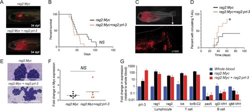
PRL-3 enhances circulation of leukemia cells in a zebrafish T-ALL model.a Representative images of transient transgenic zebrafish expressing rag2:Myc + rag2:mCherry (n = 11) or rag2:Myc + rag2:mCherry + rag2:prl-3 (n = 6) at 34 days post-fertilization (dpf). b Kaplan–Meier analysis of time (days) percent survival (>70% of animal is mCherry-positive). c Representative rag2:Myc + rag2:mCherry + rag2:prl-3 animal, showing circulating mCherry + leukemia cells within the tail fin. d Kaplan–Meier analysis of time (days) for each T-ALL to be visualized in circulation, * p = 0.049. e Representative images of May-Gunwald Giemsa staining of blood samples from fish from each leukemia type. Scale bar = 100 μm. f Realtime RT-PCR analysis of Myc expression between rag2:Myc + rag2:mCherry (n = 8) and rag2:Myc + rag2:mCherry + rag2:prl-3 (n = 5). Each point represents one fish sample. NS = not significant. g Realtime RT-PCR analysis of lymphocyte, T-cell, and B-cell specific genes. Bars are the average expression of three samples per group.
|