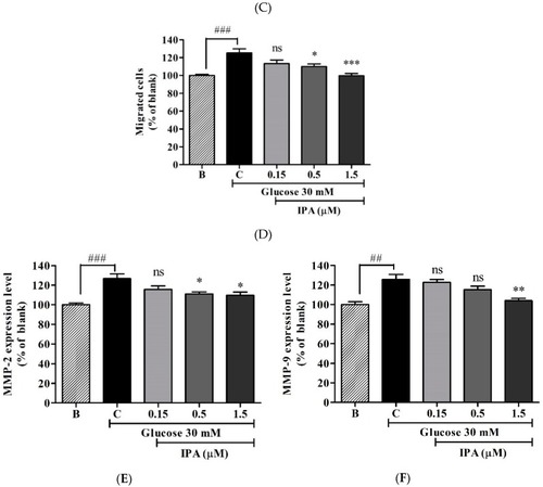
(A) IPA inhibits high glucose-induced cell migration. The cells were treated with different concentrations of IPA (0.15, 0.5, and 1.5 µM), along with 30 mM glucose. The cell monolayer was scraped at the middle of the well, and the initial gap length (0 h) and the final gap length (12 h) was measured. (B) The gap closure was quantified by percentage (%). (C) IPA inhibits cell migration through transwell filter chambers. The migrated cells were fixed, stained, and counted. (D) Quantification of migrated cells through transwell filter chambers. Matrix Metalloproteinase (MMP) (MMP-2 and -9) expression levels were evaluated using commercial Enzyme-Linked Immunosorbent Assay (ELISA) kits. (E) Quantification of MMP-2 expression. (F) Quantification of MMP-9 expression. The effects of 30 mM glucose on cell migration/MMP expression levels were compared to B (blank (0 mM glucose + 0 µM IPA)), and the effects of IPA on high glucose-induced cell migration/MMP expression level were normalized to C (control (30 mM glucose + 0 µM IPA)). Scale bar (A) and (C) 1000 µm. ns; not significant, * p ˂ 0.05, ** p ˂ 0.01, *** p ˂ 0.001, ##p ˂ 0.01, ###p ˂ 0.001.
|
