|
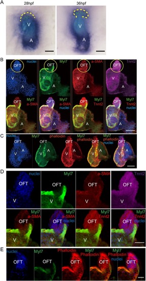
Identification of the TNNT2-positive, Myl7-negative smooth muscle cells in the OFT. (A) Images of in situ hybridization with tnnt2a anti-sense probe. Yellow dotted line, outflow tract outline; green dotted line, cell outline. (B) Immunofluorescence of Tnnt2, Myl7, and α-SMA staining in wild-type zebrafish hearts at 5 dpf. Nuclei is stained by Hoechst 33342. Yellow circle shows OFT area. (C) Myl7 zebrafish heart stained by phalloidin (F-actin) at 5 dpf. Nuclei is stained by Hoechst 33342. Scale bar: 150 µm. (D,E) Images of a single plane of Tnnt2, Myl7, α-SMA and phalloidin immunofluorescence staining. V, ventricle; A, atrium. Scale bars: (A–C) 150 µm, (D,E) 50 µm.
|