FIGURE

Figure 1

ID
ZDB-FIG-191230-1705
Publication
Brenet et al., 2019 - Defective Excitatory/Inhibitory Synaptic Balance and Increased Neuron Apoptosis in a Zebrafish Model of Dravet Syndrome
Other Figures
All Figure Page
Back to All Figure Page
Figure 1

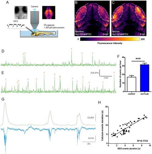
Correlation between LFP and calcium activity in scn1Lab model. (A) Schematic of the experimental setup to simultaneously record local field potential and calcium activity of a larval zebrafish brain. (B,C) Calcium images showing respectively the baseline activity (B) and the seizure activity (C). The fluorescence intensity is color-coded as shown in the color bar below the images. (D,E) Representative 20 min calcium recording of immobilized and paralyzed 4 dpf control (D) (N = 9) and scn1Lab (E) (N = 9) larvae. Each calcium event is shown with an orange arrowhead. (F) Number of events, calcium increases greater that 0.04 ΔF/F0 amplitude, during 1 h recording. (G) Representative LFP, recorded in the left neuropil of the optic tectum, and calcium traces, from the optic tectum, of a paralyzed and immobilized 4 dpf scn1Lab larva. (H) Correlation between LFP and calcium events duration (n = 46). N = number of larvae and n = number of events. Error bars on all graphs represent standard error of the mean (SEM). ***, p < 0.001. p-value was determined using Student’s unpaired t-test.

Expression Data

Expression Detail
Antibody Labeling
Phenotype Data
Fish:
Knockdown Reagent:
Observed In:
Stage: Day 4

Phenotype Detail
Acknowledgments
This image is the copyrighted work of the attributed author or publisher, and ZFIN has permission only to display this image to its users. Additional permissions should be obtained from the applicable author or publisher of the image. Full text @ Cells