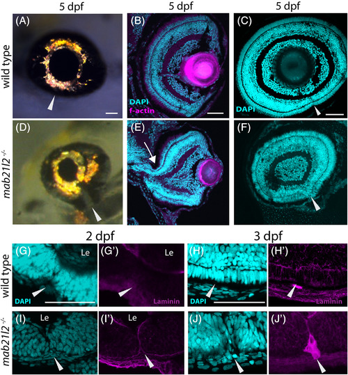
mab21l2−/− mutants possess colobomas of varying severity, and retain basement membrane markers in the choroid fissure. A,D: Whole‐mount images of wild‐type (A) and mab21l2−/− mutant (D) embryos highlighting colobomas. Compared with wild‐type, mab21l2−/− embryos show colobomas of varying severities at 5 dpf (D). B,C,E,F: Transverse (B,E) and sagittal (C,F) sections of wild‐type (B,C) and mab21l2−/− mutant (E,F) embryos at 5 dpf. Note the severe coloboma in proximal eye cup of the mab21l2−/− mutant (E, arrow) compared with wild‐type (B). In sagittal section view, (C,F), the mab21l2−/− retina (F) displays discontinuity of retinal lamina and failure of choroid fissure fusion (arrowhead) when compared with wild‐type (C). G‐J′: Laminin α1 localization in wild‐type (G,H′) and mab21l2−/− (I,J′) eyes. Magenta = laminin, cyan = DAPI. Le = lens. Arrowhead marks the site of the choroid fissure. Note that wild‐type embryos (G,G′,H,H′) do not display laminin α1 at the site of the closed choroid fissure, while mab21l2−/− mutants (I,I′,J,J′) retain laminin α1 localization at the open choroid fissure (arrowheads). Scale bars = 50 μm
This image is the copyrighted work of the attributed author or publisher, and
ZFIN has permission only to display this image to its users.
Additional permissions should be obtained from the applicable author or publisher of the image.
Full text @ Dev. Dyn.
Your Input Welcome
Thank you for submitting comments. Your input has been emailed to ZFIN curators who may contact you if
additional information is required.
Oops. Something went wrong. Please try again later.