
Fig. 7
- ID
- ZDB-FIG-190925-25
- Publication
- Gath et al., 2019 - Zebrafish mab21l2 mutants possess severe defects in optic cup morphogenesis, lens and cornea development
- Other Figures
- All Figure Page
- Back to All Figure Page
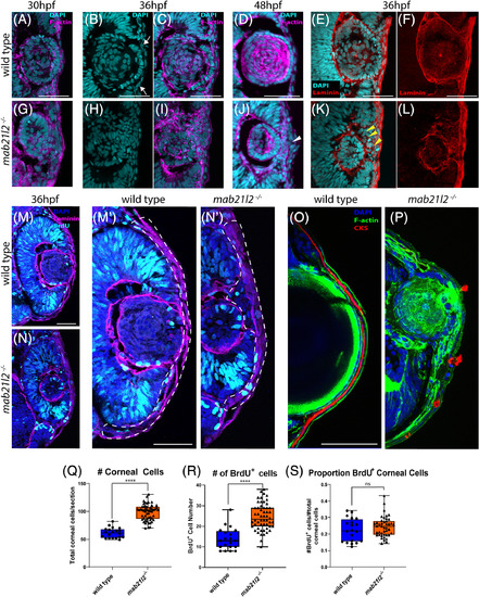
mab21l2 −/− mutants display corneal dysgenesis and failure of stromal patterning. A‐D,G‐J: Wild‐type (A‐D) and mab21l2 −/− mutant (G‐J) sections demonstrating corneal phenotypes. At 30 and 36 hpf, when compared with wild‐type eyes (A‐C), mab21l2 −/− lenses (G‐I) appear continuous with the overlying surface ectoderm. At 48 hpf, mab21l2 −/− mutants (J) possess a multilayered mass of cells (arrowhead) at the ocular surface when compared with wild‐type (D). E,F,K,L: Laminin α1 distribution in 36 hpf wild‐type (E,F) and mab21l2 −/− mutant (K,L) embryos demonstrating the presence of ectopic, nonlens, nonretinal cells in mab21l2 −/− mutants (K, arrowheads). M‐N′: BrdU incorporation assays of 36 hpf wild‐type (M) and mab21l2 −/− mutant (N) embryos. At 36 hpf, mab21l2 −/− mutants (N) possess more corneal cells, but these cells are not ectopically proliferative relative to wild‐type (M). Zooms in M′ and N′; dotted lines show area counted for quantification. O‐P: Corneal keratan sulfate (CKS) stain of 5 dpf wild‐type (O) and mab21l2 −/− mutant (P) embryos. Mutants do not properly express CKS in the stroma. Q‐S: Quantification of total and BrdU+ corneal cells in M and N. (P < 0.0001, P < 0.0001, P = 0.27, respectively). Scale bars = 50 μm |
| Fish: | |
|---|---|
| Observed In: | |
| Stage Range: | Prim-15 to Long-pec |