FIGURE
Fig. 5
- ID
- ZDB-FIG-190925-23
- Publication
- Gath et al., 2019 - Zebrafish mab21l2 mutants possess severe defects in optic cup morphogenesis, lens and cornea development
- Other Figures
- All Figure Page
- Back to All Figure Page
Fig. 5
|
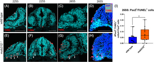
mab21l2 −/− possess elevated cell death in their optic stalk. A‐C,E‐G: TUNEL stain of wild‐type (A‐C) and mab21l2 −/− mutant (E‐G) embryos. Compared with wild‐type, 22SS mutant embryos (E,F) possess an increase in cell death in the optic stalk region of the eye. This difference persists through 26SS (G). D,H: Pax2 and TUNEL co‐stain of 26SS wild‐type (D) and mab21l2−/−(H) embryos. Compared with wild‐type (D), mab21l2 −/−embryos possess increased dying cells in the pax2+optic stalk region. I: Quantification of the proportion of pax2+ and TUNEL+ cells in D and H. Note a significantly higher (P = 0.014) proportion of pax2+ cells are TUNEL+ in mab21l2 −/− mutants when compared with wild‐type embryos. Dorsal is up in all panels. Scale bars = 50 μm |
Expression Data
| Gene: | |
|---|---|
| Fish: | |
| Anatomical Term: | |
| Stage: | 26+ somites |
Expression Detail
Antibody Labeling
Phenotype Data
| Fish: | |
|---|---|
| Observed In: | |
| Stage Range: | 20-25 somites to 26+ somites |
Phenotype Detail
Acknowledgments
This image is the copyrighted work of the attributed author or publisher, and
ZFIN has permission only to display this image to its users.
Additional permissions should be obtained from the applicable author or publisher of the image.
Full text @ Dev. Dyn.

