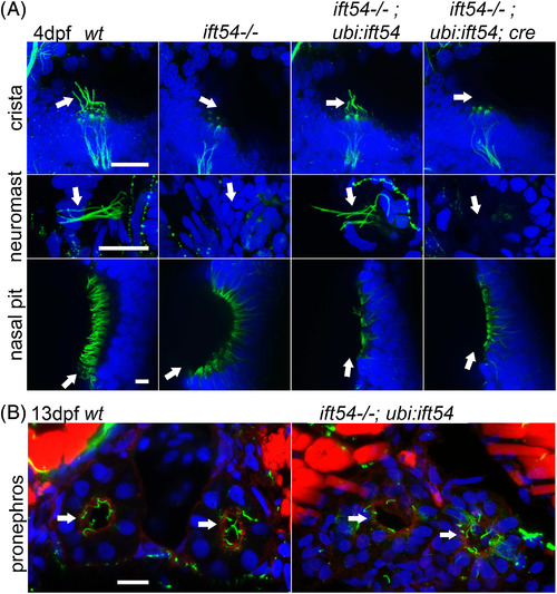
Fig. 3
|
The Tg(ubi:loxP‐ift54‐loxP‐myr‐mcherry,myl7:EGFP)sh488 rescue transgene restores cilia in ift54tp49 mutants. A: Confocal stack projections of the crista of the ear and neuromasts, and confocal slices of the nasal epithelium of 4‐dpf embryos. Acetylated tubulin immunofluorescence of cilia axonemes (green); DAPI staining of DNA in blue. Arrows show cilia axonemes or the positions where they are missing. Images shown are representative of all of the embryos examined of each group from ift54 tp4/+;Tg(ubi:loxP‐ift54‐loxP‐myr‐mcherry,myl7:EGFP)sh488 /+ x ift54 tp49/+ crosses with or without cre mRNA injection. The rescue transgene restores cilia formation in the crista and neuromasts but not nasal epithelium. B: Confocal stack projections of transverse sections of trunk at 13 dpf. Acetylated tubulin (green) immunofluorescence showing cilia axonemes of the pronephric ducts (white arrows), phalloidin staining of actin in red, and DAPI staining of DNA in blue. Images representative of all of three ift54 tp49; Tg(ubi:loxP‐ift54‐loxP‐myr‐mcherry,myl7:EGFP)sh488 fish (ift54−/−;ubi:ift54) and three wt sibs (wt) examined. The rescue transgene rescues cilia of pronephric ducts. WT, wild‐type. Scale bars = 20 μm |
| Fish: | |
|---|---|
| Observed In: | |
| Stage Range: | Day 4 to Days 7-13 |

