|
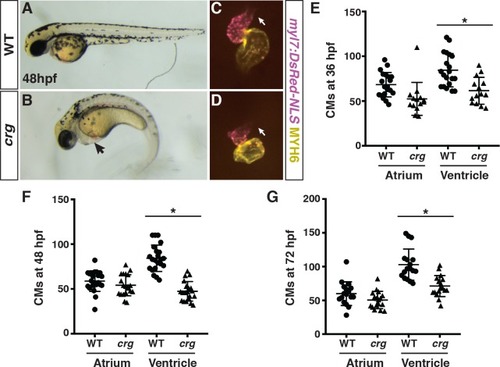
<italic>Crg</italic> mutants have a specific deficit in VCs.(A-B) WT sibling and crg mutants at 48 hpf. Lateral views with anterior to the left. Arrow in B indicates pericardial edema. (C-D) Hearts from WT sibling and crg mutant myl7:NLS-DsRed2 embryos at 48 hpf. Frontal views. Purple alone indicates ventricle. Yellow indicates atrium. Arrows indicate arterial pole of the ventricle. (E-G) Quantification of CMs in the atria and ventricles of WT sibling and crg mutant myl7:DsRed2-NLS embryos at 36, 48, and 72 hpf. For 36 hpf, n = 14 for WT and crg mutants. For 48 hpf, n = 20 for WT and crg mutants. For 72 hpf, n = 17 for WT and crg mutants. Asterisk in all graphs indicates p<0.05 as determined by Student’s t-test. Error bars for all graphs indicate s.e.m.
|