FIGURE

Fig 1

ID
ZDB-FIG-190723-156
Publication
Hartwell et al., 2019 - Anteroposterior patterning of the zebrafish ear through Fgf- and Hh-dependent regulation of hmx3a expression
Other Figures
All Figure Page
Back to All Figure Page
Fig 1

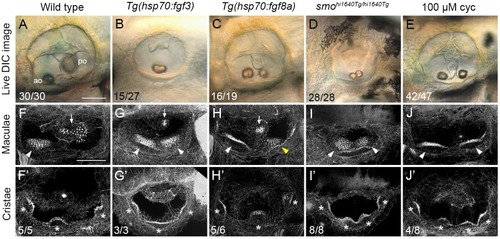
Duplicate double-anterior ear phenotypes resulting from early Fgf mis-expression or Hh pathway inhibition.

(A–E) Differential interference contrast (DIC) images of ears in live embryos at 3 dpf (72 hpf). (F–J’) Confocal images of FITC-phalloidin stains, revealing stereociliary bundles on sensory hair cells in the maculae (F–J) or cristae (F’–J’). Anterior maculae and duplicate anterior maculae are marked with arrowheads; posterior maculae and remnants of posterior maculae are marked with arrows. Cristae and duplicate cristae are marked with asterisks. Yellow arrowhead in H indicates macula that is ventromedial in position, and close to remnants of the posterior macula (arrow). Note the enlarged lateral crista in G’. (The bright spot in the centre of F’ is a lateral line neuromast.) Representative phenotypes are shown; numbers of embryos displaying these phenotypes are indicated on the panels. All Tg(hsp70:fgf3) heat-shocked embryos (n = 27) and smohi1640Tg/hi1640Tg mutants (n = 28) showed double-anterior ears. In B, 15/27 ears had a single fused otolith as shown; the remaining 12/27 ears had two separate, but small and ventrally-positioned otoliths. In C, 16/19 ears showed varying degrees of duplication with two small, ventrolaterally positioned otoliths (14/19, as shown) or fused otoliths (2/19). The remaining 3/19 ears showed a wild-type phenotype. In D, 17/28 ears had two otoliths touching as shown; in the remaining 11/28 ears, the otoliths were separate, but both small and ventrally positioned. Genotypes or treatments are indicated for each column. Transgenic lines were subject to 30 minutes of heat shock at 14 hpf. Additional controls for this figure are shown in S1 Fig. Lateral views; anterior to the left. Abbreviations: ao, anterior (utricular) otolith; po, posterior (saccular) otolith; cyc, cyclopamine. Scale bar in A, 50 μm (applies to A–E); scale bar in F, 50 μm (applies to F–J’).

Expression Data

Expression Detail
Antibody Labeling
Phenotype Data
Fish:
Conditions:
Observed In:
Stage: Protruding-mouth

Phenotype Detail
Acknowledgments
This image is the copyrighted work of the attributed author or publisher, and ZFIN has permission only to display this image to its users. Additional permissions should be obtained from the applicable author or publisher of the image. Full text @ PLoS Genet.