
Fig 1
- ID
- ZDB-IMAGE-190723-200
- Publication
- Hartwell et al., 2019 - Anteroposterior patterning of the zebrafish ear through Fgf- and Hh-dependent regulation of hmx3a expression
- All Figures
- Figures for Hartwell et al., 2019
|
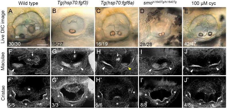
Fig 1
(A–E) Differential interference contrast (DIC) images of ears in live embryos at 3 dpf (72 hpf). (F–J’) Confocal images of FITC-phalloidin stains, revealing stereociliary bundles on sensory hair cells in the maculae (F–J) or cristae (F’–J’). Anterior maculae and duplicate anterior maculae are marked with arrowheads; posterior maculae and remnants of posterior maculae are marked with arrows. Cristae and duplicate cristae are marked with asterisks. Yellow arrowhead in H indicates macula that is ventromedial in position, and close to remnants of the posterior macula (arrow). Note the enlarged lateral crista in G’. (The bright spot in the centre of F’ is a lateral line neuromast.) Representative phenotypes are shown; numbers of embryos displaying these phenotypes are indicated on the panels. All