
Fig. 2
- ID
- ZDB-FIG-190709-3
- Publication
- Ahsan et al., 2019 - Prickle1 is required for EMT and migration of zebrafish cranial neural crest
- Other Figures
- All Figure Page
- Back to All Figure Page
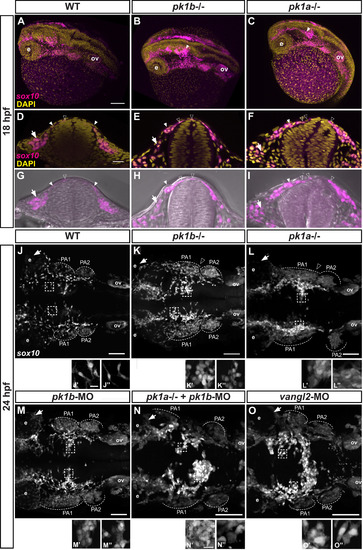
Disruption of pk1b or pk1a function disrupts cranial NCC disposition. (A-C) Maximal projections of confocal images in dorsolateral view of fixed Tg(sox10:EGFP) 18 hpf embryos (EGFP indicated in magenta), counterstained with DAPI (yellow), show the 3D formation of NCC streams migrating ventrolaterally. The pk1b fh122/ fh122 (B) and pk1ach105/ch105 (C) mutant embryos exhibit dorsal clusters of NCCs along the AP axis, as indicated by solid white arrowheads; ov = otic vesicle, e = eye, scale bar = 100 µm. (D-I) Single confocal z-slices of transverse sections through the anterior hindbrain of wild-type (D, G), pk1b fh122/ fh122 (E, H) and pk1ach105/ch105 (F, I) Tg(sox10:EGFP) 18 hpf embryos counterstained with DAPI (yellow; D-F) or together with bright-field views (G-I). Open arrowheads indicate nuclei of dorsally located NCCs (EGFP signal in magenta), closed arrowheads indicate NCCs in process of ventrolateral migration around the neural keel, arrows indicate NCCs that have migrated out into the surrounding head mesenchyme, scale bar = 25 µm. (J-O) Maximal projections of confocal images in dorsal view from fixed, deyolked, flat-mounted Tg(sox10:EGFP) embryos at 24 hpf. The scale (bar = 100 µm) is identical for all specimens shown except for pk1ach105/ch105 + pk1b-morphant, and vangl2-morphant embryos, which showed a shortened AP body axis and a wider mediolateral body axis due to convergence defects. As compared to WT embryos (n = 13 embryos) (J), pk1b fh122/ fh122 (n = 12 embryos) (K) and pk1ach105/ch105 (n = 9 embryos) (L) mutant embryos exhibit the maintenance of distinct dorsally-localized NCC clusters. pk1b-morphant (n = 18 embryos) (M) embryos phenocopy the pk1b fh122/ fh122 mutant embryos (K). The pk1ach105/ch105 + pk1b-morphant (n = 8 embryos) (N), and vangl2-morphant (n = 7 embryos) (O) show a more severe phenotype of dorsal NCC clustering across the midline. (J′-O’’) High magnification insets from associated micrographs (boxed) show that in contrast to WT specimens (J′, J’’) where NCCs exhibit bipolar, protrusive morphologies, pk1b fh122/ fh122 (K′, K’’) pk1ach105/ch105 (L′, L’’), pk1b-morphant (M′, M’’), pk1ach105/ch105 + pk1b-morphant (N′, N’’), and vangl2-morphant (O′, O’’) NCCs show less protrusive, more rounded morphologies that exhibit close contacts with neighboring NCCs. The scale (bar = 10 µm) is identical for each inset. PA1 =pharyngeal arch 1, PA2 =pharyngeal arch 2, PA3 =pharyngeal arch 3, ov=otic vesicle. |
| Gene: | |
|---|---|
| Fish: | |
| Anatomical Term: | |
| Stage: | 14-19 somites |
| Fish: | |
|---|---|
| Knockdown Reagents: | |
| Observed In: | |
| Stage Range: | 14-19 somites to Prim-5 |
Reprinted from Developmental Biology, 448(1), Ahsan, K., Singh, N., Rocha, M., Huang, C., Prince, V.E., Prickle1 is required for EMT and migration of zebrafish cranial neural crest, 16-35, Copyright (2019) with permission from Elsevier. Full text @ Dev. Biol.