
Fig. 4
- ID
- ZDB-FIG-190328-46
- Publication
- Palha et al., 2018 - An in vivo translation-reporter system for the study of protein synthesis in zebrafish embryos
- Other Figures
- All Figure Page
- Back to All Figure Page
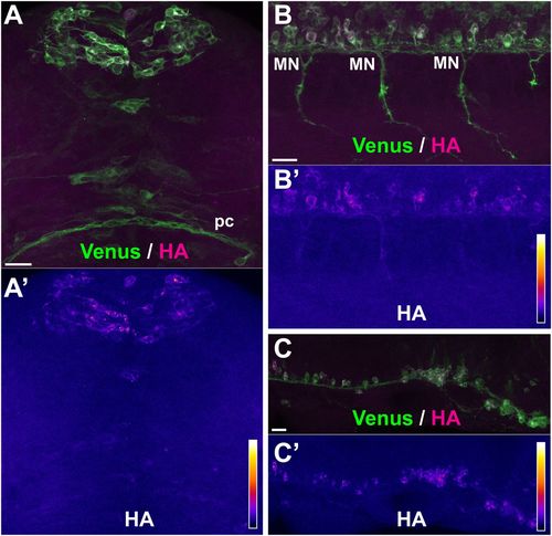
No axonal translation occurs with soma-restricted 3′UTR sequences. Representative examples of sites of protein synthesis revealed by HA/Venus immunostaining of Danoprevir-treated, SPoT transgenic embryos containing 3'UTRs with restricted axonal mRNA localization. (A–C) Superimposed Venus (green) and HA (magenta) immunostaining signals. (A′–C′) HA immunostaining signal intensity color-coded with imageJ ‘Fire’ lookup table. Color-intensity correspondence is represented in the calibration bar. (A,B) Transgenic line, Tg(SPoT_neuroD). Danoprevir treatment time=1.5 h. (C) Transgenic line, Tg(SPoT_chicken ß-actin-ΔZipcode). Danoprevir treatment time, 1.5 h. Scale bars: 20 µm. |