
Fig. 1
- ID
- ZDB-FIG-190328-43
- Publication
- Palha et al., 2018 - An in vivo translation-reporter system for the study of protein synthesis in zebrafish embryos
- Other Figures
- All Figure Page
- Back to All Figure Page
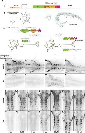
Protease-controlled translation reporter system. (A) Principle of the SPoT method. (1) The translation reporter cassette encodes a myristoylated form of the Yellow Fluorescent Protein Venus fused to NS3 protease domain, flanked by two of its own specific cleavage sites, and a HA epitope tag. (2) The UAS-controlled promoter ensures expression of the construct in neurons in Tg(huC:Gal4) transgenic line. (3) In normal conditions, the protease cleaves itself rapidly after translation, removing the HA peptide. At time T0, addition of a small cell-permeable molecule (Danoprevir) prevents protease activity and stabilizes subsequently synthesized proteins, maintaining their HA tag. HA-labeled proteins, that have been synthesized during the Δt period between T0 and fixation, reveal sites of translation. (B–E′) Flatmounted immunostaining for Venus (top row) and HA (bottom row) of Tg(SPoT_chicken ß-actin) embryos incubated for 1 h with Danoprevir (D,D′), Cycloheximide (C,C′), both (E,E′) or none (B,B′). (F–L′) Flatmounted immunostaining for Venus (top row) and HA (bottom row) of Tg(SPoT_chicken ß-actin) embryos incubated for 1.5 h with increasing concentrations of Danoprevir. Scale bars: 200 µm. |