
Fig. 6
- ID
- ZDB-FIG-181023-4
- Publication
- Yamaguchi et al., 2018 - Systematic studies of all PIH proteins in zebrafish reveal their distinct roles in axonemal dynein assembly
- Other Figures
- All Figure Page
- Back to All Figure Page
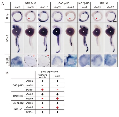
DNAH genes showed distinct expression patterns in zebrafish embryos and testis. (A) Whole-mount in situ hybridization of DNAH genes with embryos (12 and 32 hpf) and dissected testis (seminal lobule). For embryos, lateral views are shown. Yolk of 12 hpf embryos was removed before observation to show Kupffer’s vesicle clearly. Red arrowheads indicate Kupffer’s vesicles or testes in which DNAH gene expressions were not detected. White asterisks: non-specific staining of yolk. Scale bars: 200 μm for embryos; 100 μm for testes. (B) Comparison of DNAH gene expression between Kupffer’s vesicle and testis. +, expressed; -, expression not detected. Red indicates when DNAH gene expression was difference between the two organs. |
| Genes: | |
|---|---|
| Fish: | |
| Anatomical Terms: | |
| Stage Range: | 5-9 somites to Adult |