
FIGURE
Fig. 7-S1
- ID
- ZDB-FIG-181023-5
- Publication
- Yamaguchi et al., 2018 - Systematic studies of all PIH proteins in zebrafish reveal their distinct roles in axonemal dynein assembly
- Other Figures
- All Figure Page
- Back to All Figure Page
Fig. 7-S1
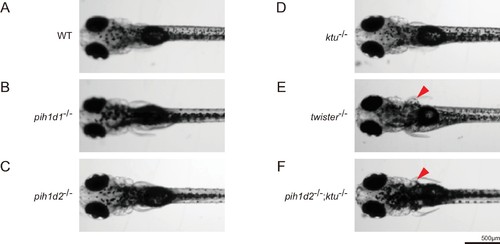
Expansion of pronephric duct was observed in twister-/- and pih1d2-/-;ktu-/-. (A–F) Dorsal views of zebrafish larvae at 4 days post-fertilization. (A) WT, (B) pih1d1-/-, (C) pih1d2-/-, (D) ktu-/-, (E) twister-/-, (F) pih1d2-/-;ktu-/-. twister-/- and pih1d2-/-;ktu-/- exhibited abnormal expansions of pronephric ducts (red arrowheads), which disabled larvae from having their pectoral fins close to their bodies. |
Expression Data
Expression Detail
Antibody Labeling
Phenotype Data
Phenotype Detail
Acknowledgments
This image is the copyrighted work of the attributed author or publisher, and
ZFIN has permission only to display this image to its users.
Additional permissions should be obtained from the applicable author or publisher of the image.
Full text @ Elife