
Fig. S5
- ID
- ZDB-FIG-181019-44
- Publication
- Yin et al., 2018 - Spatiotemporal Coordination of FGF and Shh Signaling Underlies the Specification of Myoblasts in the Zebrafish Embryo
- Other Figures
- All Figure Page
- Back to All Figure Page
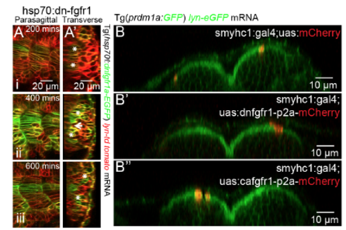
SSF migration under global or local FGFR perturbation, related to Figure 6 (A) Time lapse of slow muscle migration under heat shock of hs:dnfgfr1-GFP. (A’) reconstructed transverse view from (A). White asterisks label the migrating SSFs. Images are taken at muscle segments 18-21. (B) Lateral migration remains intact under mosaic FGF perturbations in slow muscles driven by smyhc1:gal4;UAS:dnfgfr1-p2a-mCherry (B’) and smyhc1:gal4;UAS:cafgfr1-p2a-mCherry (B’’) compared with smyhc1:gal4;UAS:mCherry as a control (B). Lateral to the top and dorsal to the right. Images are taken at muscle segments 14-16 at 30hpf. |
Reprinted from Developmental Cell, 46, Yin, J., Lee, R., Ono, Y., Ingham, P.W., Saunders, T.E., Spatiotemporal Coordination of FGF and Shh Signaling Underlies the Specification of Myoblasts in the Zebrafish Embryo, 735-750.e4, Copyright (2018) with permission from Elsevier. Full text @ Dev. Cell