
Fig. S4
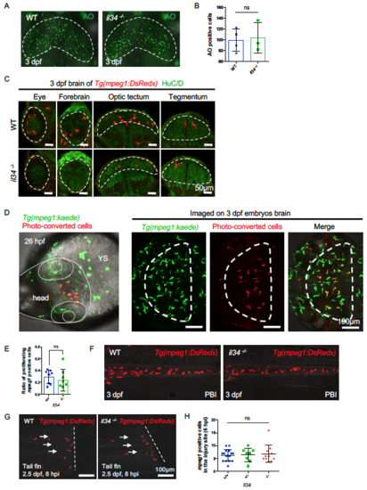
Characterization of il34 Mutant Embryos. Related to Figure 3. (A and B) Acridine orange (AO) staining showing apoptotic neurons (A) and quantification (B) of apoptotic neurons in the optic tectum of 3dpf WT or il34-/- embryos injected with pu.1 morpholino. The optic tectum is indicated by dashed lines. n = 4 and 3 for WT and il34-/- embryos, respectively. Values represent means with SD. (C) Transverse sections showing microglia in different CNS regions of 3 dpf WT or il34-/- embryos, with macrophages labeled by Tg(mpeg1:DsRedx) (red) and neurons labeled by HuC/D immunostaining (green). Different CNS regions are indicated by dashed lines. Related to Figure 3E. (D) 10 Kaede+ (before-convert: green) macrophages in the anterior head region of 26 hpf Tg(mpeg1:kaede) embryos were photo-converted (after-convert: red). Images taken at 3 dpf showing the successful colonization of converted macrophages (red) in the optic tectum. The head is indicated by dotted lines and the optic tectum is indicated by dashed lines. YS, yolk sac. (E) Quantification of the ratio of proliferating DsRedx+ macrophages in total head macrophages in the time-lapse imaging of WT Tg(mpeg1:DsRedx) or il34-/-; Tg(mpeg1:DsRedx) embryos from 30 hpf to 34 hpf. n = 8 for each group. Values represent means ± SD. (F) Representative images of macrophages in the PBI region of 3 dpf WT or il34-/- embryos. Macrophages were labeled by Tg(mpeg1:DsRedx) (red). (G and H) Representative images (left) and quantification (right) of macrophages accumulating in the injury sites 6 hours after injuries performed in the tail fins of 2 dpf il34+/+ (n = 16), il34+/- (n = 8) and il34-/- (n = 11) embryos. Macrophages were labeled by Tg(mpeg1:DsRedx) (red). Values represent means ± SD. ns, P>0.05. |
| Fish: | |
|---|---|
| Knockdown Reagent: | |
| Observed In: | |
| Stage: | Protruding-mouth |
Reprinted from Developmental Cell, 46, Wu, S., Xue, R., Hassan, S., Nguyen, T.M.L., Wang, T., Pan, H., Xu, J., Liu, Q., Zhang, W., Wen, Z., Il34-Csf1r Pathway Regulates the Migration and Colonization of Microglial Precursors, 552-563.e4, Copyright (2018) with permission from Elsevier. Full text @ Dev. Cell