
Fig. 2
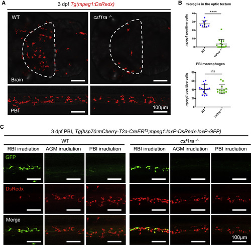
csf1ra Deficiency Blocks the Colonization of Microglia but Not Peripheral Macrophages (A and B) Representative images (A) and quantification (B) of macrophages in the brain (upper panel) or the PBI (lower panel) of 3-dpf WT or csf1ra−/− embryos. Macrophages were labeled by Tg(mpeg1:DsRedx) (red). The optic tectum is indicated by dashed lines. n = 9, 15, 15, and 13 for WT microglia, csf1ra−/− microglia, WT PBI macrophages, and csf1ra−/− PBI macrophages, respectively. Values represent means ± SD. (C) Double-transgenic Tg(hsp70:mCherry-T2a-CreERT2;mpeg1:loxP-DsRedx-loxP-GFP) WT or csf1ra−/− embryos were irradiated using an infrared (IR) laser in the RBI, AGM, and PBI region at 14 hpf, 28 hpf, and 22 hpf, respectively, to induce expression of CreER. After 4-hydroxytamoxifen (4-OHT) treatment, CreER mediates loxP recombination and results in GFP expression. Images show the GFP+ and DsRedx+ macrophages in the PBI region of 3-dpf irradiated embryos in the respective group. ns, p > 0.05; ∗∗∗∗, p ≤ 0.0001. |
| Gene: | |
|---|---|
| Fish: | |
| Anatomical Terms: | |
| Stage: | Protruding-mouth |
| Fish: | |
|---|---|
| Observed In: | |
| Stage: | Protruding-mouth |
Reprinted from Developmental Cell, 46, Wu, S., Xue, R., Hassan, S., Nguyen, T.M.L., Wang, T., Pan, H., Xu, J., Liu, Q., Zhang, W., Wen, Z., Il34-Csf1r Pathway Regulates the Migration and Colonization of Microglial Precursors, 552-563.e4, Copyright (2018) with permission from Elsevier. Full text @ Dev. Cell