
FIGURE
Fig. S6
- ID
- ZDB-FIG-180918-31
- Publication
- Xing et al., 2018 - Mutational analysis of dishevelled genes in zebrafish reveals distinct functions in embryonic patterning and gastrulation cell movements
- Other Figures
- All Figure Page
- Back to All Figure Page
Fig. S6
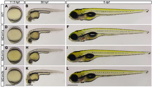
Phenotypes of MZdvl1a, MZdvl1b and MZdvl3b mutants at different stages. (A-C) WT embryos. (D-F) MZdvl1a mutants. (G-I) MZdvl1b mutants. (J-L) MZdvl3b mutants. All embryos are lateral view. The anterior region of 11.5 hpf embryos is positioned on the top. Scale bars: (A, D, G, J) 400 μm; (B, E, H, K) 400 μm; (C, F, I, L) 400 μm. |
Expression Data
Expression Detail
Antibody Labeling
Phenotype Data
Phenotype Detail
Acknowledgments
This image is the copyrighted work of the attributed author or publisher, and
ZFIN has permission only to display this image to its users.
Additional permissions should be obtained from the applicable author or publisher of the image.
Full text @ PLoS Genet.