
Fig. 3
- ID
- ZDB-FIG-180918-24
- Publication
- Xing et al., 2018 - Mutational analysis of dishevelled genes in zebrafish reveals distinct functions in embryonic patterning and gastrulation cell movements
- Other Figures
- All Figure Page
- Back to All Figure Page
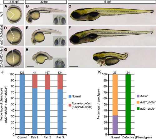
Dvl2 and Dvl3a dosages in axis extension and AP patterning. Both dvl2+/-;MZdvl3a and Zdvl2;MZdvl3a mutants were from crosses between dvl2+/-;dvl3a-/- carriers. Representative mutant embryos were imaged at indicated stages. Eye phenotypes are shown in the insets as ventral view. (A-C) WT embryos. (D-F) dvl2+/-;MZdvl3a mutants display moderate axis extension defect at 11.5 hpf and 30 hpf, and recover to a normal phenotype at 5 dpf. (G-I) Zdvl2;MZdvl3a mutants show strong axis extension defect at different stages, and display caudal truncation, craniofacial defects, cardiac edema (arrow), and fused eyes or cyclopia (inset) at 30 hpf and at 5 dpf. (J) Quantitative analysis of the posterior truncation phenotype at 5 dpf in offspring derived from three independent dvl2+/-;dvl3a-/- carriers. Control embryos were from crosses between female WT fish and male dvl2+/-;dvl3a-/- fish. Posterior deficiency is present in the offspring from all three fish pairs with a proportion that follows the Mandel inheritance (about 25%). Numbers on the top of each column indicate total embryos analyzed. (K) Genotyping of normal and posteriorly truncated embryos. All defective embryos are dvl2-/-;dvl3a-/- mutants. Scale bars: (A, D, G) 400 μm; (B, E, H) 400 μm; (C, F, I) 400 μm. |
| Fish: | |
|---|---|
| Observed In: | |
| Stage Range: | 1-4 somites to Day 5 |