
Fig. 1
- ID
- ZDB-FIG-180918-22
- Publication
- Xing et al., 2018 - Mutational analysis of dishevelled genes in zebrafish reveals distinct functions in embryonic patterning and gastrulation cell movements
- Other Figures
- All Figure Page
- Back to All Figure Page
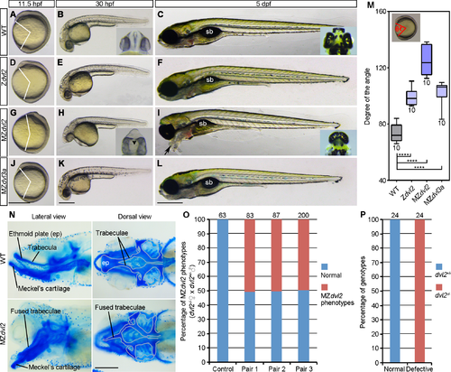
Analysis of dvl2 and dvl3a mutant phenotypes. (A-C) WT embryos, the insets show the eyes in ventral view at 30 hpf and in dorsal view at 5 dpf. (D-F) Zdvl2 mutants, obtained by crosses between heterozygous dvl2+/- carriers, display weak axis extension defect at 11.5 hpf. They are normal at 30 hpf, and show reduced swim bladder (sb) at 5 hpf. (G-I) MZdvl2 mutants, obtained from crosses between female dvl2-/- fish and male dvl2+/- fish, display a reduced AP axis at 11.5 hpf and 30 hpf. They present fused eyes at 30 hpf (inset, ventral view; arrowhead indicates fused lenses), and develop craniofacial defects and cyclopia (inset, dorsal view), with pharyngeal cartilages protruding outward (arrow) at 5 dpf. (J-L) MZdvl3a mutants from crosses between dvl3a-/- carriers display weak axis extension defect, but are indistinguishable from WT embryos at 30 hpf and 5 dpf. (M) Statistical analysis of the extent of axis extension delay. The embryos were imaged at 11.5 hpf followed by genotyping. Those embryos with expected genotypes were used to measure the angle between the anterior end and posterior end, with vertex at the geometric center of the embryo (inset). Bars represent the mean ± s.d. from indicated numbers of embryos (****, P<0.0001). (N) Alcian blue staining of head cartilages at 5 dpf. Cartilage structures of the basicranium are outlined in grey, showing the fusion of trabeculae and the absence of ethmoid plate (ep) in MZdvl2 mutants. (O) Quantitative analysis of MZdvl2 mutant phenotypes at 5 dpf in offspring from three independent female dvl2-/- and male dvl2+/- fish pairs. Control embryos were obtained from crosses between female WT fish and male dvl2+/- fish. Numbers on the top of each column indicate total embryos analyzed. (P) Genotyping of dvl2 mutants with normal and defective phenotypes. All embryos with a normal phenotype are dvl2+/- mutants, whereas all defective embryos are MZdvl2 mutants. Numbers on the top of each column indicate total embryos genotyped from three independent fish pairs. Scale bars: (A, D, G, J) 400 μm; (B, E, H, K) 400 μm; (C, F, I, L) 400 μm; (N) 100 μm. |
| Fish: | |
|---|---|
| Observed In: | |
| Stage Range: | 1-4 somites to Day 5 |