
Fig. S2
- ID
- ZDB-FIG-180627-32
- Publication
- Flanagan-Steet et al., 2018 - TGF-ß Regulates Cathepsin Activation during Normal and Pathogenic Development
- Other Figures
- All Figure Page
- Back to All Figure Page
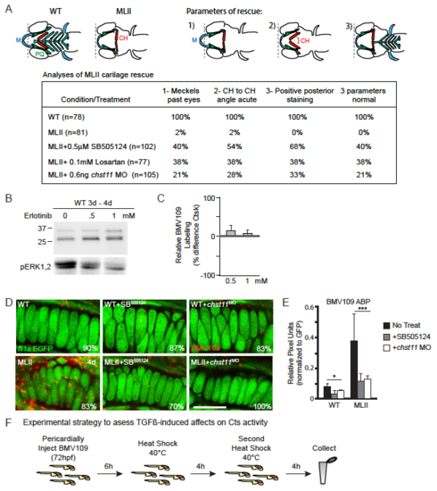
TGFß modulation impacts Ctsk activity and MLII phenotypes. A) Strategy for quantitating rescue in Alcian blue stained embryos. Schematic shows cartilage morphology in WT versus MLII embryos. M=Meckel’s cartilage, CH=ceratohyal. In MLII, the ceratohyals do not extend beyond the eye (dotted line), the angle between ceratohyal is typically close to 180°C, and the posterior structures (inlcuding the pectoral fin) do not stain. Rescue is assessed as the numberof embryos with parameters 1,2,3 all restored. The table details the percent of embryos with each individual parameter rescued, as well as the percent of embryos with all three parameters normal. B)Treatment with the EGFR-inhibitor erlotinib does not alter the degree of BMV109 labeled cathepsin activity. C) Quantitation of differences in the level of BMV109 labled Ctsk in drug treated versus untreated embryos from 4 independent experiments. Error=SD.* p<0.05, **p<0.01, ***p<0.001 D,E) Confocal analyses of BMV109-labeled activity (red) in EGFP marked chondrocytes shows Cts actvity is locally reduced after bothTGFß inhibition (SB505124) and chst11 knockdown. F) Experimental strategy for heat shock induction of TGFß signaling. |