
Fig. 3
- ID
- ZDB-FIG-180608-29
- Publication
- van Leeuwen et al., 2018 - A transgenic zebrafish model for the in vivo study of the blood and choroid plexus brain barriers using claudin 5.
- Other Figures
- All Figure Page
- Back to All Figure Page
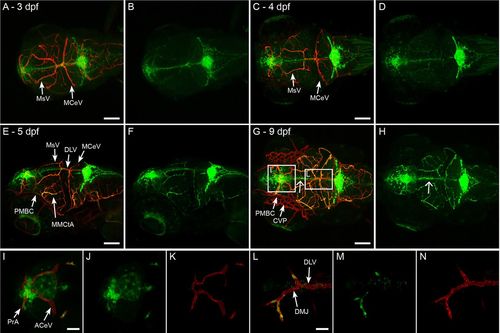
Development of Claudin 5a expression in brain vasculature. (A,B) Dorsal view of head of Tg(kdrl:mCherry)is5;TgBAC(cldn5a:EGFP)vum2 larva at 3 dpf. Merge of both channels is shown in A, single green fluorescent expression is shown in B. Claudin 5a is first expressed in the mesencephalic vein (MsV) and middle cerebral vein (MCeV). (C-F) Expansion of claudin 5a expression over time, with almost all blood vessels expressing claudin 5a at 5 dpf (E-F): dorsal view (C,D), dorsal/lateral view (E,F). (G,H) Dorsal view of head of Tg(kdrl:mCherry)is5;TgBAC(cldn5a:EGFP)vum2 larva at 9 dpf. The boxed areas are shown enlarged in I-K and L-N. Open arrow shows the strong expression of claudin 5a in the midline in the absence of a colocalizing blood vessel (H). Specific spots that never express claudin 5a are the primordial midbrain channels (PMBC) and choroidal vascular plexus (CVP) (G), anterior cerebral vein (ACeV) (I), and dorsal midline junction (DMJ) and dorsal longitudinal vein (DLV) (L). Scale bars: 100 μm in A,C,E,G; 25 μm in I,L. |
| Genes: | |
|---|---|
| Fish: | |
| Anatomical Terms: | |
| Stage Range: | Protruding-mouth to Days 7-13 |