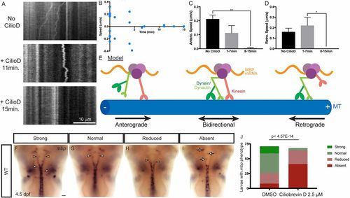
Acute dynein inhibition arrests both anterograde and retrograde Mbp mRNA transport in cultured oligodendrocytes and perturbs mbp localization in zebrafish. (A) In kymographs representing 60 s of live-cell imaging, MS2-GFP–labeled Mbp mRNA can be seen moving in the retrograde direction in untreated cells (Top). Following acute ciliobrevin D (15 μM) addition, Mbp mRNA displays bidirectional motility characterized by frequent back-and-forth movement and many directional switches (Middle). Finally, around 15 min after ciliobrevin D (15 μM) addition, most cells display arrested motility (Bottom). (B) A scatter plot represents the average anterograde or retrograde net speeds for individual oligodendrocytes. (C and D) Anterograde or retrograde net speeds were averaged for each cell and binned across 27-min time periods following ciliobrevin treatment. At 8–15 min following ciliobrevin treatment, speeds significantly decreased compared with earlier time points. One-way ANOVAs with post hoc Tukey’s test were performed. Anterograde speeds: No CilioD vs. 1–7 min, P = 0.11; No CilioD vs. 8–15 min, **P = 0.0011; 1–7 min vs. 8–15 min, P = 0.077. Retrograde speeds: No CilioD vs. 1–7 min, P = 0.67; No CilioD vs. 8–15 min, P = 0.11, 1–7 min vs. 8–15 min, *P = 0.027. (E) A model shows that Mbp mRNA granules can move processively in the anterograde and retrograde directions as well as bidirectionally and that each Mbp mRNA granule can simultaneously bind to both kinesin and the dynein/dynactin complex. (F–I) mbp ISH of zebrafish larvae treated with ciliobrevin D for 21 h. Larvae were scored as having strong (F), normal (G), reduced (H), or absent (I) mbp localization in oligodendrocyte processes (arrowheads, processes; arrows, cell bodies). (J) Quantification of phenotypic distribution shows that there was a significant difference in scores for DMSO (n = 71) and ciliobrevin D-treated larvae (n = 68), with the latter exhibiting more larvae with reduced or absent mbp localization in oligodendrocyte processes (P < 4.5E-14, Fisher’s exact test). (Scale bar, 50 μM.)
|
