
Fig. 7
- ID
- ZDB-FIG-180320-10
- Publication
- Suarez-Bregua et al., 2017 - Targeted Pth4-expressing cell ablation impairs skeletal mineralization in zebrafish
- Other Figures
- All Figure Page
- Back to All Figure Page
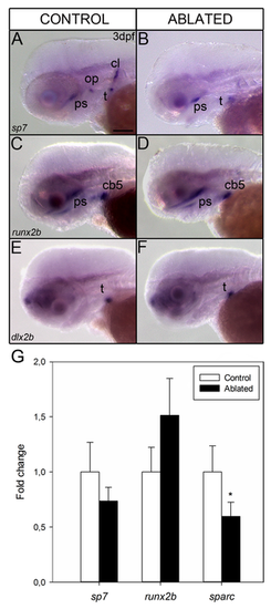
Whole mount in situ hybridization shows the gene expression pattern of osteoblast differentiation markers at 3 dpf in control (A, C and E) and ablated larvae (B, D and F) from lateral view. Expression of sp7 is detected in operculum, cleithrum, teeth and parasphenoid (A). Note the absence of expression in cleithrum and operculum in ablated larvae (B). runx2b and dlx2b display the same expression pattern before ablation (C, E) and after ablation (D, F). Quantitative RT-PCR analysis of early (runx2b), intermediate (sp7) and late (sparc) stage markers of osteoblast differentiation in 7 dpf ablated (black bars) and control larvae (white bars) (G). Results show significant down-regulation of sparc gene expression (*p <0.05) when compared with controls. Results normalized to actb1 are expressed as mean ± SEM. Abbreviations: ps, parasphenoid; op, operculum; cb5, ceratobranchial arch 5; t, teeth; cl, cleithrum. Scale bars: 100 μm. |
| Genes: | |
|---|---|
| Fish: | |
| Condition: | |
| Anatomical Terms: | |
| Stage Range: | Protruding-mouth to Days 7-13 |
| Fish: | |
|---|---|
| Condition: | |
| Observed In: | |
| Stage Range: | Protruding-mouth to Days 7-13 |