
FIGURE
Fig. 2
- ID
- ZDB-FIG-180320-6
- Publication
- Suarez-Bregua et al., 2017 - Targeted Pth4-expressing cell ablation impairs skeletal mineralization in zebrafish
- Other Figures
- All Figure Page
- Back to All Figure Page
Fig. 2
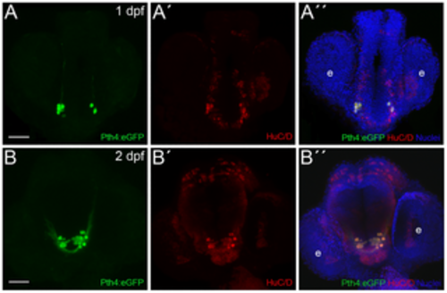
Zebrafish Pth4:eGFP-expressing cells are post-mitotic neurons. Double inmunostaining in Pth4:eGFP transgenic embryos using anti-eGFP antibody (A and B) and anti-HuC/D antibody (A´and B´) shows complete co-localization at 1 and 2 dpf (A´´ and B´´). Ventral views with anterior to the top. Pth4:eGFP: green; HuC/D: red; nuclear stain: blue. Abbreviation: e, eye. Scale bar: 50 μm. |
Expression Data
| Antibody: | |
|---|---|
| Fish: | |
| Anatomical Term: | |
| Stage Range: | Prim-5 to Long-pec |
Expression Detail
Antibody Labeling
Phenotype Data
Phenotype Detail
Acknowledgments
This image is the copyrighted work of the attributed author or publisher, and
ZFIN has permission only to display this image to its users.
Additional permissions should be obtained from the applicable author or publisher of the image.
Full text @ PLoS One