
FIGURE
Fig. 3
- ID
- ZDB-FIG-180207-2
- Publication
- Hale et al., 2017 - Shp2-MAPK signalling drives proliferation during zebrafish embryo caudal fin-fold regeneration
- Other Figures
- All Figure Page
- Back to All Figure Page
Fig. 3
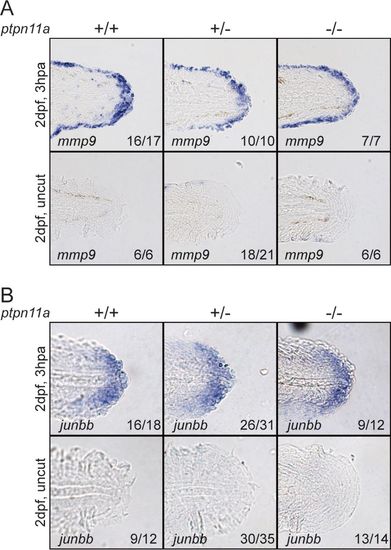
Formation of wound epidermis and distal blastema in zebrafish embryos lacking functional Shp2. At 2 dpf, the caudal fin folds of zebrafish embryos from a ptpn11a+/− ptpn11b−/− incross were amputated and allowed to regenerate. The embryos were fixed at 3 hpa, or the equivalent for uncut controls, and subjected to hybridization using probes specific for mmp9 (A) or junbb (B). Representative images of caudal fin folds of genotyped embryos are shown, and the number of embryos showing similar patterns/total number of embryos analyzed are indicated in the bottom right corner of each image. |
Expression Data
| Genes: | |
|---|---|
| Fish: | |
| Condition: | |
| Anatomical Terms: | |
| Stage: | Day 5 |
Expression Detail
Antibody Labeling
Phenotype Data
Phenotype Detail
Acknowledgments
This image is the copyrighted work of the attributed author or publisher, and
ZFIN has permission only to display this image to its users.
Additional permissions should be obtained from the applicable author or publisher of the image.
Full text @ Mol. Cell. Biol.