
Fig. 1
- ID
- ZDB-FIG-180207-1
- Publication
- Hale et al., 2017 - Shp2-MAPK signalling drives proliferation during zebrafish embryo caudal fin-fold regeneration
- Other Figures
- All Figure Page
- Back to All Figure Page
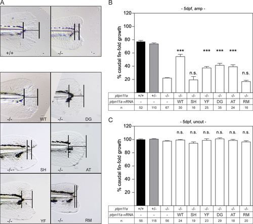
Functional Shp2a is required for regeneration. Zebrafish embryos from a ptpn11a+/− ptpn11b−/− incross were microinjected at the one-cell stage with synthetic mRNA encoding wild-type Shp2a, SH2 domain mutant Shp2a-R32M-R138M, C-terminal tyrosine mutant Shp2a-Y542F-Y580F, Shp2a-D61G, Shp2a-A462T, or Shp2a-R466M or were not injected (−). At 2 dpf, the caudal fin fold was amputated, and regeneration was assessed at 3 dpa (i.e., 5 dpf and 3 dpa); equivalent uncut controls were included (i.e., 5 dpf, uncut). All the embryos were genotyped. (A) Representative images of amputated ptpn11a−/− ptpn11b−/− embryo caudal fin folds at 3 dpa. A ptpn11a+/+ ptpn11b−/− sibling in which regeneration of the caudal fin fold was 80% complete by 3 dpa is shown for comparison (top left). (B and C) Regeneration was quantified by measuring the distance from the tip of the notochord to the edge of the caudal fin fold, as indicated (bars in panel A). The means of caudal fin fold growth are depicted relative to caudal fin fold growth of uncut ptpn11a+/+ ptpn11b−/− controls. Means of microinjected amputated (amp) (B) or uncut (C) ptpn11a−/− ptpn11b−/− embryos were compared to those of noninjected amputated or uncut ptpn11a−/− ptpn11b−/− embryos. The data were pooled from multiple experiments. Statistical evaluation was performed using a Mann-Whitney U test for comparison of ptpn11a−/− ptpn11b−/− zebrafish embryos with siblings within amputated or uncut groups, not between amputated and uncut groups. The error bars indicate standard errors of the mean. ***, P < 0.001; n.s. not significant. |
| Fish: | |
|---|---|
| Condition: | |
| Observed In: | |
| Stage: | Day 5 |