
Fig. 2
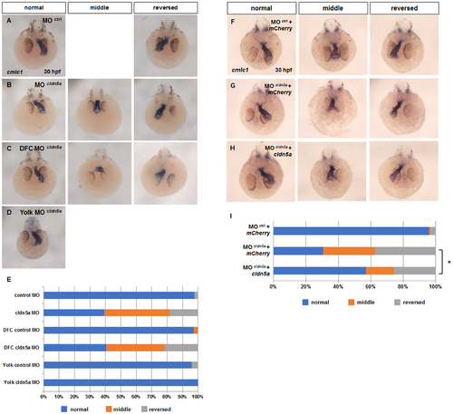
Laterality of heart was disrupted in cldn5a morphants. (A—H) Visualization of a heart by in situ hybridization of cmlc1 in 30 hpf embryos. Representative images of control morphants (A), cldn5a morphants (B), DFC cldn5a morphants (C), and yolk cldn5a morphants (D). (E) Stacked bar graph (blue; normal, orange; middle, grey; reversed, control morphants; n = 45, cldn5a morphants; n = 33, DFC control morphants; n = 71, DFC cldn5a morphants; n = 37, yolk control morphants; n = 25, yolk cldn5a morphants; n = 31). Representative images of control morphants with mCherry (F), cldn5a morphants with mCherry (G), cldn5a morphants with mCherry-cldn5a (H). (I) Stacked bar graph (blue; normal, orange; middle, grey; reversed, control morphants with mCherry; n = 132, cldn5a morphants with mCherry; n = 111, cldn5a morphants with mCherry-cldn5a; n = 115). * depicts p < 0.05. |
| Gene: | |
|---|---|
| Fish: | |
| Knockdown Reagent: | |
| Anatomical Term: | |
| Stage: | Prim-15 |
| Fish: | |
|---|---|
| Knockdown Reagent: | |
| Observed In: | |
| Stage: | Prim-15 |