
Fig. 5
- ID
- ZDB-FIG-180104-5
- Publication
- Djenoune et al., 2017 - The dual developmental origin of spinal cerebrospinal fluid-contacting neurons gives rise to distinct functional subtypes
- Other Figures
- All Figure Page
- Back to All Figure Page
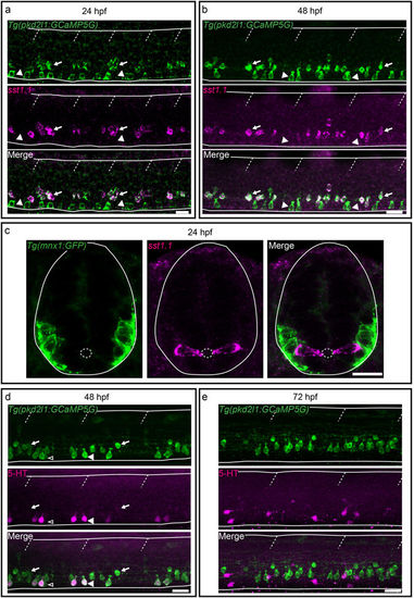
Secreted factors distinguish dorsal and ventral CSF-cNs: while dorsal express the somatostatin paralog sst1.1, ventral CSF-cNs express 5-HT. (a–c) Lateral views of the spinal cord show that sst1.1 expression is restricted to dorsal CSF-cNs (arrows, FISH for sst1.1 (magenta) coupled to GFP IHC (green) on Tg(pkd2l1:GCaMP5G)) (a,b) and Tg(mnx1:GFP) (c) embryos and larvae at 24 hpf (a,c) and 48 hpf (b). (c) Transverse sections show that sst1.1 (magenta) is not expressed in motor neurons (green) as previously suggested (Devos et al.52). (d,e) IHC for 5-HT (magenta) and GFP (green) on Tg(pkd2l1:GCaMP5G) transgenic larvae at 48 hpf (d) and 72 hpf (e). (d) At 48 hpf, most ventral CSF-cNs express 5-HT (arrowhead, compared to negative cells shown with empty arrowhead). Note that dorsal CSF-cNs (arrows) are not labelled by 5-HT. (e) At 72 hpf, ventral CSF-cNs are not serotoninergic anymore in the rostral part of the spinal cord. Horizontal lines represent the limits of the spinal cord and slash dashed lines represent somite boundaries. Small dotted ellipses represent the limit of the central canal. Scale bars = 20 μm. |
| Genes: | |
|---|---|
| Fish: | |
| Anatomical Terms: | |
| Stage Range: | Prim-5 to Protruding-mouth |