
Fig. S2
- ID
- ZDB-FIG-180104-7
- Publication
- Djenoune et al., 2017 - The dual developmental origin of spinal cerebrospinal fluid-contacting neurons gives rise to distinct functional subtypes
- Other Figures
- All Figure Page
- Back to All Figure Page
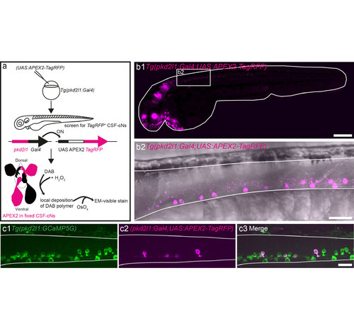
Use of the APEX2 peroxidase combined with the UAS-Gal4 system and pkd2l1 specificity to target CSF-cNs. (a) Schematic showing how APEX2 is used to generate diaminobenzidine (DAB) contrast for EM in zebrafish CSF-cNs taking advantage of the UAS-Gal4 system. We injected the (UAS:APEX2- TagRFP) construct into Tg(pkd2l1:Gal4)icm10 fertilized eggs in order to get specific expression of APEX2 in CSF-cNs. After screening for TagRFP expression, larvae were fixed and bathed with a solution of DAB and H2O2. APEX2 catalyzes the polymerization and local deposition of DAB, which subsequently attracts electron-dense osmium (OsO4), giving EM contrast only in APEX2 expressing structures namely CSF-cNs (ventral and dorsal). (b1) Lateral view of a Tg(pkd2l1:Gal4)icm10 larva at 2.5 dpf injected with (UAS:APEX2-TagRFP) showing that pkd2l1 promoter drives APEX2-TagRFP expression in CSF-cNs within the spinal cord. (b2) Close up of the rostral spinal cord from a lateral view in the same transgenic animal showing the TagRFP expression in CSF-cNs. (c) Co-injection of (pkd2l1:Gal4) and (UAS:APEX2-TagRFP) within Tg(pkd2l1:GCaMP5G)icm07 embryos where all CSF-cNs are labeled (green) demonstrates the variegated expression of TagRFP within CSF-cNs (magenta). Note that in this panel, the expression of TagRFP is restricted in some dorsal CSF-cNs. Scale bars: 300 μm (b1) and 20 μm (b2, c). |