
FIGURE
Fig. S1
- ID
- ZDB-FIG-180104-6
- Publication
- Djenoune et al., 2017 - The dual developmental origin of spinal cerebrospinal fluid-contacting neurons gives rise to distinct functional subtypes
- Other Figures
- All Figure Page
- Back to All Figure Page
Fig. S1
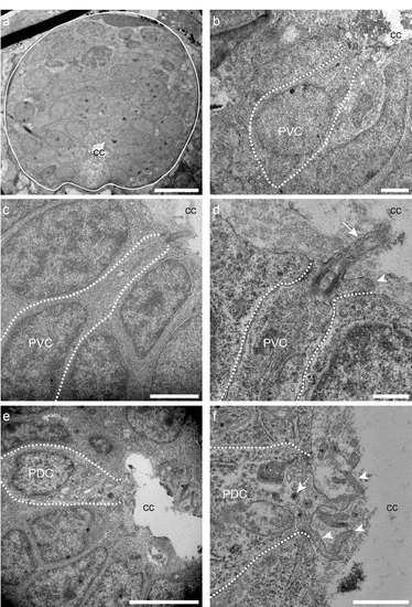
Ultrastructure of putative spinal CSF-cNs in wild type zebrafish larva. (a) Transverse section of the spinal cord illustrating multiple cells around the central canal (cc). (b-d) Putative ventral CSF-cNs referred to as PVC. (d) PVC bearing microvilli (arrowhead) and extending a cilium (arrow) with it apical pole. (e, f) Putative dorsal CSF-cN referred to as PDC extending microvilli (arrowheads) in contact with the central canal (cc). Scale bars: 10 μm (a), 2 μm (b, c), 500 nm (d), 5 μm (e) and 1 μm (f). |
Expression Data
Expression Detail
Antibody Labeling
Phenotype Data
Phenotype Detail
Acknowledgments
This image is the copyrighted work of the attributed author or publisher, and
ZFIN has permission only to display this image to its users.
Additional permissions should be obtained from the applicable author or publisher of the image.
Full text @ Sci. Rep.