
Fig. 2
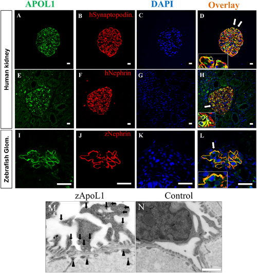
APOL1 expression in human and zebrafish glomeruli. The expression of APOL1 in podocytes is shown by double staining with an antibody against APOL1 and an antibody against the podocyte-specific proteins synaptopodin and nephrin (A-H). The regions in panel D and H labeled by arrows are shown in higher magnifications (insets). Similar to the human kidney, zApoL1 colocalizes with zNephrin in the zebrafish glomerulus (L). Panel M shows the localization of zApoL1 in podocytes (red circles) and in endothelial cells (green circles) by immunoelectron microscopy. Panel N shows the control reaction (secondary antibody only). Scale bars represent 20 μm (A-L) and 1 μm (M-N). |
| Genes: | |
|---|---|
| Antibody: | |
| Fish: | |
| Anatomical Terms: | |
| Stage: | Day 4 |