
Fig. 2
- ID
- ZDB-IMAGE-171113-50
- Genes
- Antibodies
- Publication
- Kotb et al., 2016 - Knockdown of ApoL1 in Zebrafish Larvae Affects the Glomerular Filtration Barrier and the Expression of Nephrin
- All Figures
- Figures for Kotb et al., 2016
|
Fig. 2
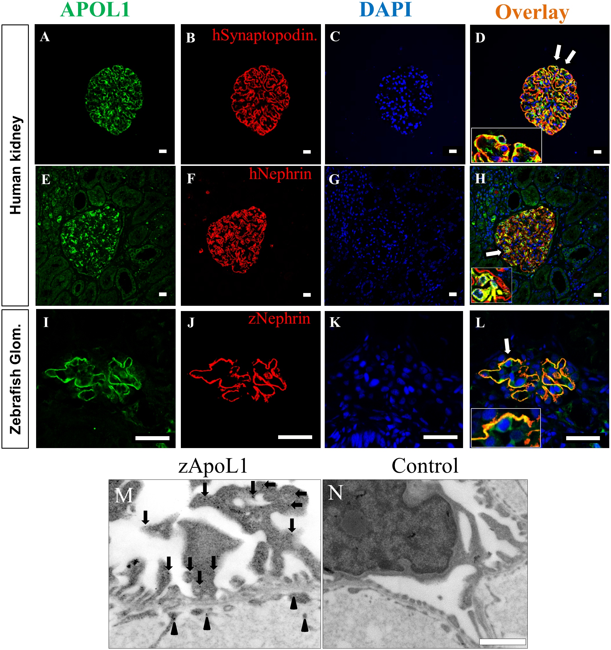
APOL1 expression in human and zebrafish glomeruli.
The expression of APOL1 in podocytes is shown by double staining with an antibody against APOL1 and an antibody against the podocyte-specific proteins synaptopodin and nephrin (A-H). The regions in panel D and H labeled by arrows are shown in higher magnifications (insets). Similar to the human kidney, zApoL1 colocalizes with zNephrin in the zebrafish glomerulus (L). Panel M shows the localization of zApoL1 in podocytes (red circles) and in endothelial cells (green circles) by immunoelectron microscopy. Panel N shows the control reaction (secondary antibody only). Scale bars represent 20 μm (A-L) and 1 μm (M-N).