
Fig. 7
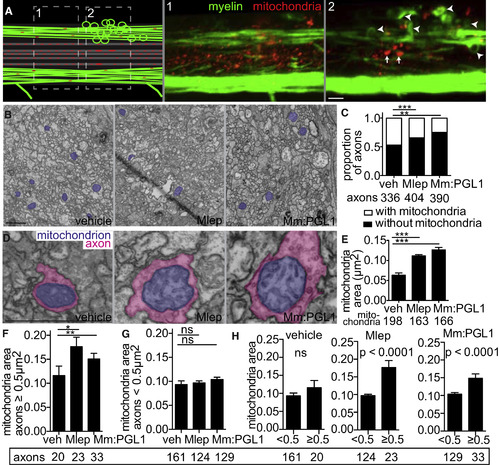
M. leprae Infection Damages Axonal Mitochondria (A) Diagram (left) of a demyelinating lesion in a 2 dpi (5 dpf) mbp larva with fluorescent mitochondria in axons. Dashed boxes indicate insets 1 and 2, with corresponding confocal images, showing mitochondria outside the lesion (inset 1) and those within the lesion (inset 2). Arrowheads indicate myelin protrusions; arrows indicate enlarged mitochondria. Scale bar, 10 μm. (B) Representative TEMs of matched anatomical regions showing the number of axon mitochondria (purple) in larvae injected with PBS, M. leprae, or M. marinum:PGL-1. Scale bar, 1 μm. (C) Proportion of axons with mitochondria to those without, in larvae like in (B); the number of axons scored are listed below (contingency analysis corrected for multiple comparisons; ∗∗p = 0.004; ∗∗∗p < 0.0002). (D) Representative TEMs of enlarged mitochondria (purple) within enlarged axons (pink), in larvae like in (B). Scale bar, 1 μm. (E) Mean (±SEM) area of mitochondria in axons, in larvae like in (B); number of mitochondria scored are listed below. (∗∗∗p < 0.001; one-way ANOVA with Dunnett’s multiple comparison.) (F) Mean (±SEM) area of mitochondria in nonmyelinated axons with area ≥0.5 μm2, in larvae like in (B) (∗p < 0.05, ∗∗p < 0.01; one-way ANOVA with Dunnett’s multiple comparison). (G) Mean (±SEM) area of mitochondria in nonmyelinated axons with area <0.5 μm2, in larvae like in (B) (one-way ANOVA with Dunnett’s multiple comparison). (H) Data from (F) and (G) are displayed per experimental group, showing mean (±SEM) area of mitochondria in large versus small nonmyelinated axons (one-way ANOVA with Dunnett’s multiple comparison). |
| Fish: | |
|---|---|
| Condition: | |
| Observed In: | |
| Stage: | Day 5 |
Reprinted from Cell, 170, Madigan, C.A., Cambier, C.J., Kelly-Scumpia, K.M., Scumpia, P.O., Cheng, T.Y., Zailaa, J., Bloom, B.R., Moody, D.B., Smale, S.T., Sagasti, A., Modlin, R.L., Ramakrishnan, L., A Macrophage Response to Mycobacterium leprae Phenolic Glycolipid Initiates Nerve Damage in Leprosy, 973-985.e10, Copyright (2017) with permission from Elsevier. Full text @ Cell