
FIGURE
Fig. S1
Fig. S1
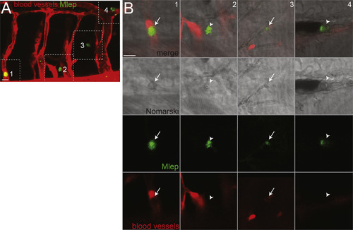
M. leprae-Infected Macrophages Escape Circulation, Related to Figures 1G and 1H (A) Confocal image from Figure 1G of a 4 dpf kdrl:dsRed larva, which has fluorescent blood vessels, 2 days post-infection (dpi) with fluorescent M. leprae; dashed lines define insets (1-4) shown in B. 10 μm bar. (B) Monochannel and merged images of insets from A, showing M. leprae within cells, likely macrophages, which are visible by Nomarski microscopy. Arrows, intracellular M. leprae retained in vessels; arrowheads, intracellular M. leprae outside vessels. 10 μm bar. |
Expression Data
Expression Detail
Antibody Labeling
Phenotype Data
Phenotype Detail
Acknowledgments
This image is the copyrighted work of the attributed author or publisher, and
ZFIN has permission only to display this image to its users.
Additional permissions should be obtained from the applicable author or publisher of the image.
Reprinted from Cell, 170, Madigan, C.A., Cambier, C.J., Kelly-Scumpia, K.M., Scumpia, P.O., Cheng, T.Y., Zailaa, J., Bloom, B.R., Moody, D.B., Smale, S.T., Sagasti, A., Modlin, R.L., Ramakrishnan, L., A Macrophage Response to Mycobacterium leprae Phenolic Glycolipid Initiates Nerve Damage in Leprosy, 973-985.e10, Copyright (2017) with permission from Elsevier. Full text @ Cell