FIGURE

Fig. 7

ID
ZDB-FIG-170606-25
Publication
Yoo et al., 2017 - Mind Bomb-Binding Partner RanBP9 Plays a Contributory Role in Retinal Development
Other Figures
All Figure Page
Back to All Figure Page
Fig. 7

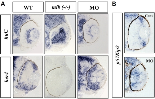
Molecular characterization of retinal development in ranbp9 morphants.

(A) Whole-mount in situ hybridization analysis for the expression of neural markers; huC for neuronal differentiation marker and her4 for neural progenitor marker, respectively. The neurogenic mutant mind bomb (mib, −/−) was used for comparison. In the WT larvae at 48 hpf, huC was expressed in the apical cells of the retina, but her4 was expressed in the basal cells. In the mib mutant larvae, huC was seen stained in most of the retinal cells near the apico-basal axis, but her4-positive cells were not observed. In the ranbp9 morphant larvae, huC expression domain was significantly reduced, however, the her4 expression domain was expanded. (B) Altered-regulation of p57kip2, a molecular marker for cell cycle exit, in ranbp9 morphants. p57kip2 transcripts were observed in the peripheral marginal regions of wild-type retina, while its expression was expanded to the central region of retina in ranbp9 morphants.

Expression Data
Genes:
Fish:
Knockdown Reagent:
Anatomical Terms:
Stage: Long-pec

Expression Detail
Antibody Labeling
Phenotype Data
Fish:
Knockdown Reagent:
Observed In:
Stage: Long-pec

Phenotype Detail
Acknowledgments
This image is the copyrighted work of the attributed author or publisher, and ZFIN has permission only to display this image to its users. Additional permissions should be obtained from the applicable author or publisher of the image. Full text @ Mol. Cells