
Fig. S10
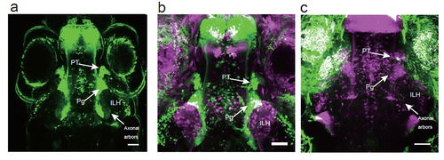
Projection of the 119B-pretectal cells to the ILH a, Merged image of 3 optical sections (66.7 μm and 22 μm apart on the Z-axis in the ventral direction) depicting the pretectal area (PT), the preglomerular nuclei (Pg), and the axonal arbours of the pretectal cells that reached the ILH at 5 dpf. Scale bar: 50 μm. b, Merged image of a UAS:EGFP;gSAIzGFFM119B larva (green) and a UAS:EGFP;hspGFFDMC76A larva (magenta, a different individual), which shows the location of the ILH relative to the PT and Pg. Scale bar: 50 μm. c, A single pretectal (PT) cell labelled by a UAS:EGFP DNA injection (green) into a UAS:RFP;gSAIzGFFM119B background zebrafish (magenta) at the 1-cell stage and observed at 4 dpf. An axon is extending out of the soma of the labelled cell in the pretectal area and is projecting to the caudomedial ILH. Scale bar: 50 μm. |