
Fig. S1
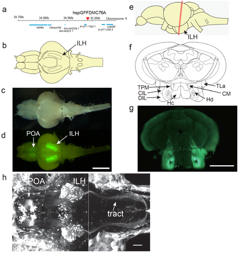
UAS:EGFP reporter gene expression in the hspGFFDMC76A Gal4 line a, Insertion site of the hspGFFDMC76A was identified in an intergenic region on chromosome 9. b. Schematic of a ventral view of the brain of an adult zebrafish. c, Ventral view of a dissected brain of an adult UAS:EGFP;hspGFFDMC76A fish. d, EGFP fluorescence in the brain shown in b. Scale bar: 1 mm. e, Schematic side view of the adult brain. The red line indicates the positions of the sections in f and g. f, Annotations based on Wulliman et al., 1996. g, EGFP fluorescence of a coronal section of the brain of an adult UAS:EGFP;hspGFFDMC76A zebrafish. Scale bar: 0.5 mm. h, UAS:EGFP expression in the hspGFFDMC76A Gal4 line at 5 dpf. Projected z-stack images obtained by two-photon laser microscopy. Left: z-stack projection of 155 slices (1 μm-step). Right: z-stack projection of 135 slices (1 μm-step). The positions of the focal planes of the two z-stacks overlap by 35 um (the right one more dorsal) along z-axis. Scale bar: 50 μm. CIL, central nucleus of the inferior lobe; CM, corpus mamillare; DIL, diffuse nucleus of the inferior lobe; Hc, caudal zone of periventricular hypothalamus; Hd, dorsal zone of periventricular hypothalamus; ILH, inferior lobes of the hypothalamus; POA, preoptic area; TLa, torus lateralis; TPM, tractus pretectomamillaris. |