
Fig. S2
- ID
- ZDB-FIG-170419-9
- Publication
- Ki et al., 2017 - Overexpression of PDGFRA cooperates with loss of NF1 and p53 to accelerate the molecular pathogenesis of malignant peripheral nerve sheath tumors
- Other Figures
- All Figure Page
- Back to All Figure Page
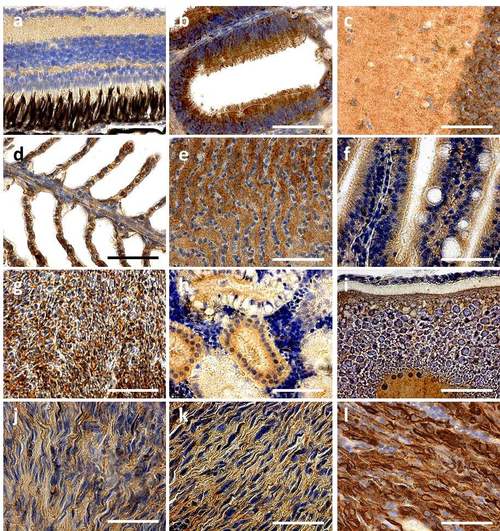
PDGFRA expressed tissues in adult fish (a-i) Representative images of PDGFRA expression by immunohistochemistry in sox10:mCherry control transgenic zebrafish in the nf1a+/-;nf1b-/- ; p53 m/m background (>30 weeks post fertilization). PDGFRA is expressed in (a) the eye inner and outer plexiform layers, (b) olfactory ciliated columnar epithelium, (c) brain, (d) gill, (e) the gill pseudobranch, (f) intestine, (g) liver, (h) kidney, and (i) vitellogenic oocytes. (j-l) Representative images of PDGFRA expression by sox10:mCherry control (j), PDGFRA wild-type (k) and mutant MPNSTs (l) in the the nf1a+/-;nf1b-/- ; p53 m/m background, respectively (>30 weeks post fertilization, scale bar = 10 μm). |