
Fig. 1
- ID
- ZDB-FIG-170419-1
- Publication
- Ki et al., 2017 - Overexpression of PDGFRA cooperates with loss of NF1 and p53 to accelerate the molecular pathogenesis of malignant peripheral nerve sheath tumors
- Other Figures
- All Figure Page
- Back to All Figure Page
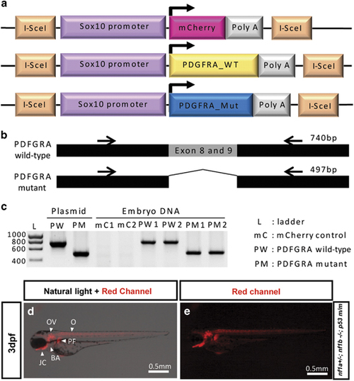
I-SceI meganuclease-mediated human PDGFRA transgenesis in nf1- and p53-deficient fish. (a) Schematic diagram of the DNA constructs used to generate transgenic sox10:mCherry, sox10:wild-type (WT) and constitutively activated (Mut) PDGFRAs zebrafish. I-SceI denotes the I-SceI meganuclease target sequence. (b) Schematic diagram of PCR target of human wild type and constitutively activated mutant PDGFRA (Δ exons 8 and 9) for genotyping. Black arrows represent primer target sites for PCR. (c) PDGFRA wild-type and mutant DNA sequences were detected in genomic DNA of the transgenic zebrafish embryos. Human PDGFRA sequences were confirmed with embryonic DNA of two separate transgenic lines (mC1 and mC2, PW1 and PW2 and PM1 and PM2). Each amplified PCR band of PDGFRA wild type and mutant had sizes of 740 and 497 bp, respectively. Injected plasmids for transgenesis were used as the positive control. (d and e) Sox10 promoter driving mCherry is expressed in cells of neural crest origin during early embryogenesis. Tg (nf1a+/−; nf1b−/−; p53m/m; sox10:mCherry) zebrafish embryo at 3 d.p.f. showed mCherry expression throughout otic vesicle (OV), branchial arches (BA), oligodendrocytes (O), jaw cartilage (JC) and pectoral fins (PF). |
| Gene: | |
|---|---|
| Fish: | |
| Anatomical Terms: | |
| Stage: | Protruding-mouth |