
Fig. 5
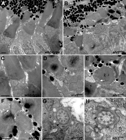
Ultrastructural analysis of wild-type and ahi1lri46 photoreceptors. (A, B) Transmission electron microscopy provided ultrastructural views of wild-type and ahi1lri46 photoreceptors. Mutant outer segments are short, fragmented, and disorganized. (C) Higher magnification of wild-type photoreceptor showing tightly stacked disc membranes and inner segment. (D) Vesiculation at the proximal end of an outer segment in ahi1lri46 mutant (arrowhead). (E) Improper orientation of disc membranes. In this field, an axoneme (white arrow) and disc membranes from outer segments of three different cells, labeled 1 to 3, are oriented perpendicular to each other. (F) Several layers of discs are abnormally wrapped around the outer segment (white arrow). (G, H) Cross-sections through the ciliary pocket (cp) show the proper 9 + 0 microtubule arrangement of axoneme. Mitochondria (m) are visible in (G). Y-shaped crosslinkers also are present (arrows in [G, H]). Staining artifacts are visible as diffuse, black deposits in some panels. Scale bars: 1 μm (A–F) and 200 nm (G, H). |
| Fish: | |
|---|---|
| Observed In: | |
| Stage: | Day 5 |