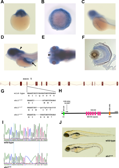
Generation of ahi1 mutant zebrafish. (A–C) Whole-mount in situ hybridization from 1 cell (A), 10 hpf (B), and 48 hpf (C), showing expression of ahi1. (D, E) Lateral and ventral views showing 5 dpf zebrafish stained by whole-mount in situ hybridization. ahi1 expression was observed throughout the central nervous system (arrowheads) and pronephros (arrow). (F) A transverse section shows ahi1 expression throughout the retina at 5 dpf. (G) Illustration of ahi1 genomic architecture. The wild-type sequence and TALENs induced ahi1 mutant alleles are shown. Wild-type nucleotides are in upper case. Deleted nucleotides are denoted as dashes while inserted nucleotides are in lower case letters. (H) Schematic protein structure of Ahi1 illustrating the location of the coiled-coil (CC) domain, WD40 repeats, and SH3 domain. The ahi1lri46 is a frame shift mutation and encodes a truncated protein lacking the 7 WD 40 repeats and the SH3 domain. (I) Chromatograms of Sanger sequencing reactions of ahi1 wild type (wild-type) and homozygous mutant (ahi1lri46) zebrafish. (J) Lateral view of representative wild-type and ahi1lri46 homozygous mutant at 5 dpf.
|
