
Fig. 3
- ID
- ZDB-FIG-170209-39
- Publication
- Noack Watt et al., 2016 - The Roles of RNA Polymerase I and III Subunits Polr1c and Polr1d in Craniofacial Development and in Zebrafish Models of Treacher Collins Syndrome
- Other Figures
- All Figure Page
- Back to All Figure Page
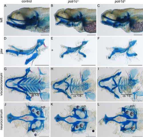
Craniofacial cartilage development is disrupted in polr1c-/- and polr1d-/- mutant embryos. (A-C) Alcian blue staining reveals cranial cartilage in 5 dpf polr1c-/- and polr1d-/- mutant embryos is hypoplastic compared to controls. (D-F) The jaws of mutant embryos are smaller overall, with noticeable differences in the size of Meckel’s cartilage, the palatoquadrate, and ceratohyal elements. (G-I) Staining of the viscerocranium reveals smaller cartilage elements derived from each of the pharyngeal arches in mutant embryos, most notably the ceratobranchials, as well as altered polarity of the ceratohyal. (J-L) Staining of the neurocranium reveals hypoplasia of the ethmoid plate. Abbreviations: M, Meckel’s cartilage; pq, palatoquadrate; ch, ceratohyal; cb, ceratobranchial; ep, ethmoid plate; pch, parachordal. Scale bar = 200 μm. |
| Fish: | |
|---|---|
| Observed In: | |
| Stage: | Day 5 |