FIGURE
Fig. S1
- ID
- ZDB-FIG-170206-7
- Publication
- Stratman et al., 2017 - Mural-Endothelial cell-cell interactions stabilize the developing zebrafish dorsal aorta
- Other Figures
- All Figure Page
- Back to All Figure Page
Fig. S1
|
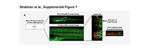
Mounting and imaging strategy of mural cell transgenic lines. (A) Embryos were mounted in 0.9% low melt point (LMP) agarose at the desired time point of imaging. Confocal stacks were acquired through the zebrafish at a 1.5 μM step size. Typically due to limitations of penetrance of lasers with confocal microscopy, one side of the embryo (the side closest to the light source) is imaged in greater detail. Excess slices from imaging are trimmed to only include the slices that include the dorsal aorta. It is key for trimming for the embryo to have been mounted as flat as possible. |
Expression Data
Expression Detail
Antibody Labeling
Phenotype Data
Phenotype Detail
Acknowledgments
This image is the copyrighted work of the attributed author or publisher, and
ZFIN has permission only to display this image to its users.
Additional permissions should be obtained from the applicable author or publisher of the image.
Full text @ Development

