
Fig. S9
- ID
- ZDB-FIG-170206-15
- Publication
- Stratman et al., 2017 - Mural-Endothelial cell-cell interactions stabilize the developing zebrafish dorsal aorta
- Other Figures
- All Figure Page
- Back to All Figure Page
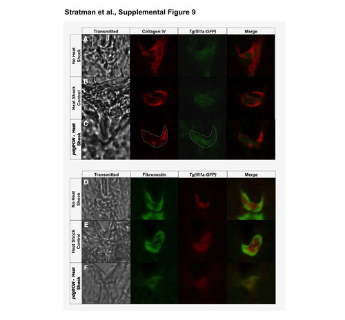
Co-staining of vascular and basement membrane proteins shows vascular enrichment. (A-C) Collagen IV with vascular co-immunostaining: column 1- transmitted light image of the region of interest; column 2- collagen IV immunolabeling (pseudocolored red); column 3- immunolabeling of GFP expressed by the Tg(fli1a:GFP) zebrafish line (pseudocolored green); column 4- merge of the collagen IV and vascular images. Three conditions were analyzed: No Heat Shock Controls (A), Heat Shock Controls (B), and pdgfrDN- Heat Shock (C). (D-F) Fibronectin with vascular co-immunostaining: column 1- transmitted light image of the region of interest; column 2- fibronectin immunolabeling (pseudocolored green); column 3- immunolabeling of GFP expressed by the Tg(fli1a:GFP) zebrafish line (pseudocolored red); column 4- merge of the collagen IV and vascular images. Three conditions were analyzed: No Heat Shock Controls (D), Heat Shock Controls (E), and pdgfrDN- Heat Shock (F). Representative images were chosed from 3 independent rounds of immunostaining. |